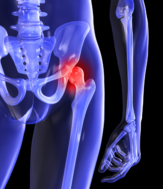
Femoroacetabular impingement (FAI) is a multifactorial hip disease in which pain is a direct result of a collision of femoral head (the ball of the hip joint) with the edge of the acetabula (the socket of the hip joint). The hallmark of the disease is pain provoked by hip flexion. Although impingement occurs during flexion of the hip joint such as with sitting, squatting and lifting the hip. Once the impingement is established the pain may appear on walking, kicking and sporting activities involving body rotation over the planted leg. This occurs because the hip joint labrum becomes damaged and the capsule becomes inflamed. The joint space may not become decreased until the progression of the disease. Labral tears may or may not be present with FAI.
Most common location of pain is in the groin, however it may appear on the side or the back of the hip.
FAI is a disease of young athletic adults. However, hip impingement also occurs in middle age population as well and account for 10-15 % of adults with any hip diseases. In older adults the changes in the hip joint occur earlier but may manifest later in life.
There are 2 types of FAI; Cam and Pincer deformity. However majority of cases are of a mixed type.CAM type – is a widening of the femoral neck. Pincer type is when there is an outgrowth of the edge of the bony acetabula.

ANATOMY Hip joint is anatomically the most complex joint in the human body. It is a part of functional unit called lumbopelvic-hip complex and as such plays a decisive role in mobility of this complex .The intrinsic stability of this joint is provided by the labrum and the capsule-ligamentous complex. The labrum is a fibrocartilagenous pad which smoothes the surface of the acetabula providing adherence and congruency to the movement of the femoral head (the ball) in the acetabula (the socket).
The articular surface of the joint is layered with hyaline cartilage- which functions to decrease friction and helps to increase range of motion. The anatomy of the hip is designed in such a way to create negative pressure between the surface of the femur and the acetabula. With Femoroacetabular impingement this pressure is lost.
Patient history and clinical examination are sufficient for diagnosing FAI. Diagnostic ultrasound may show impingements, labrum tears and associated soft tissue damage. However, MRI is much more accurate for determining the stage and extent of FAI. If surgery is not being considered, a combination of Xray and ultrasound is enough to guide rehab and/or injection therapy.
Although arthroscopic techniques have been dramatically improved and there is much more research data in the last five years the first treatment of choice is certainly conservative. Research shows that 85% of patients with Femoroacetabular hip impingement will improve with physical therapy.Research also shows that those patients with FAI who prehabed have better outcomes after the surgery.