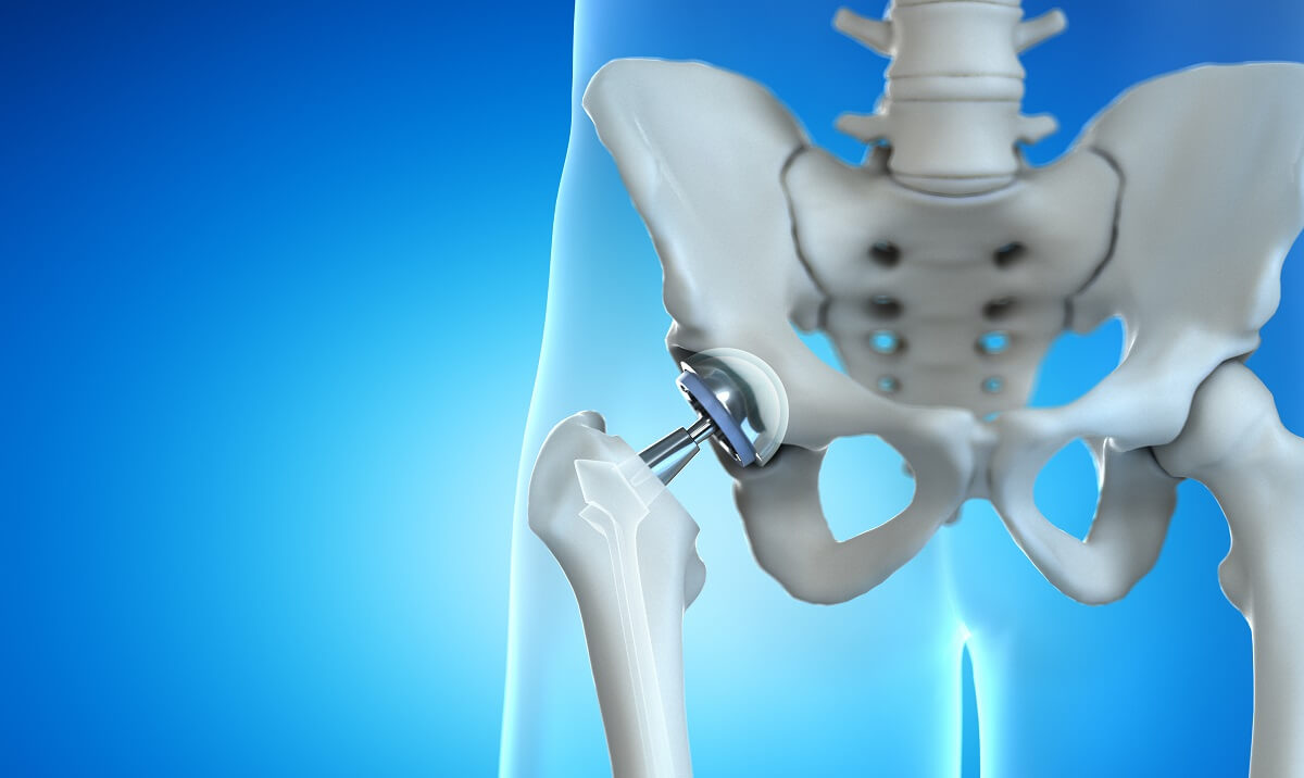
A hip replacement, or hip arthroplasty, is surgery that replaces a diseased or injured hip joint by with an artificial joint or implant. People usually receive hip replacements when arthritis causes severe hip pain and inflammation. Hip fracture and natural wear-and-tear are also common reasons for hip replacements surgery.
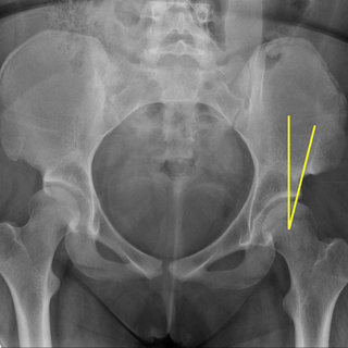
Hip arthroscopy is a surgical procedure that allows doctors to view the hip joint without making a large incision (cut) through the skin and other soft tissues. Arthroscopy is used to diagnose and treat a wide range of hip problems. During hip arthroscopy, your surgeon inserts a small camera, called an arthroscope, into your hip joint. The camera displays pictures on a video monitor, and your surgeon uses these images to guide miniature surgical instruments.
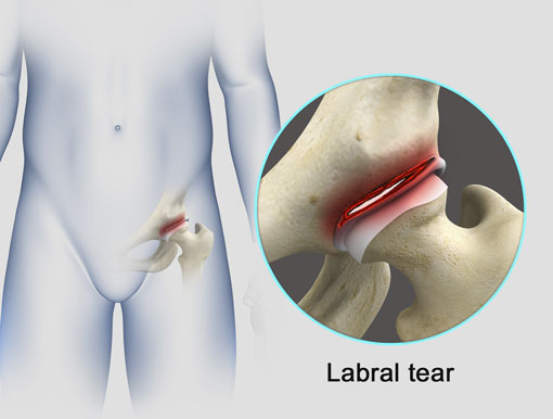
A hip labral tear involves the ring of cartilage (labrum) that follows the outside rim of your hip joint socket. Besides cushioning the hip joint, the labrum acts like a rubber seal or gasket to help hold the ball at the top of your thighbone securely within your hip socket.
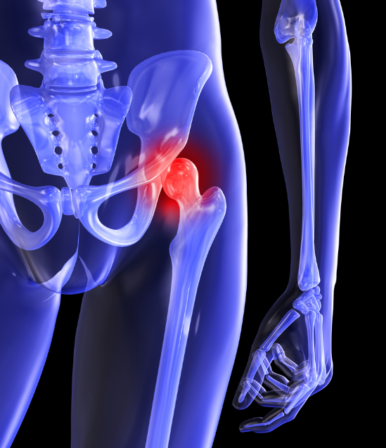
Femoroacetabular impingement (FAI) is a multifactorial hip disease in which pain is a direct result of a collision of femoral head (the ball of the hip joint) with the edge of the acetabula (the socket of the hip joint). The hallmark of the disease is pain provoked by hip flexion. Although impingement occurs during flexion of the hip joint such as with sitting, squatting and lifting the hip.
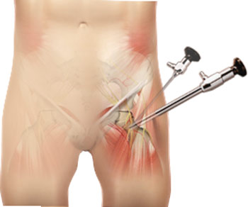
Hip arthroscopy is surgery that is done by making small cuts around your hip and looking inside using a tiny camera. Other medical instruments may also be inserted to examine or treat your hip joint. Hip arthroscopy is a minimally invasive surgical procedure performed through 2 or 3 small 5-10 mm incisions (i.e., key hole surgery), using an advanced HD camera and special instruments to visualise and work inside and around the hip joint.